Sunday, March 13, 2011
Saturday, March 12, 2011
algebraic expression
ALGEBRAIC EXPRESSIONS
An algebraic expression is made up of the signs and symbols of algebra. These symbols include the Arabic numerals, literal numbers, the signs of operation, and so forth. Such an expression represents one number or one quantity. Thus, just as the sum of 4 and 2 is one quantity, that is, 6, the sum of c and d is one quantity, that is, c + d. Likewise , ab, a - b, and so forth, are algebraic expressions each of which represents one quantity or number.
Longer expressions may be formed by combinations of the various signs of operation and the other algebraic symbols, but no matter how complex such expressions are they still represent one number. Thus the algebraic expression is one number
The arithmetic value of any algebraic expression depends on the values assigned to the literal numbers. For example, in the expression 2x2 - 3ay, if x = -3, a = 5, and y = 1, then we have the following:
2x2 - 3ay = 2(-3)2 -3(5)(1)
= 2(9) - 15 = 18 - 15 = 3
Notice that the exponent is an expression such as 2x2 applies only to the x. If it is desired to indicate the square of 2x, rather than 2 times the square of x, then parentheses are used and the expression becomes (2x)2.
Practice problems. Evaluate the following algebraic expressions when a = 4, b = 2, c = 3, x = 7, and y = 5. Remember, the order of operation is multiplication, division, addition, and subtraction.
Answers:
1. 53
2. -29
3. 19
4. 53
TERMS AND COEFFICIENTS
The terms of an algebraic expression are the parts of the expression that are connected by plus and minus signs. In the expression 3abx + cy - k, for example, 3abx, cy, and k are the terms of the expression.
An expression containing only one term, such as 3ab, is called a monomial (mono means one). A binomial contains two terms; for example, 2r + by. A trinomial consists of three terms. Any expression containing two or more terms may also be called by the general name, polynomial (poly means many). Usually special names are not given to polynomials of more than three times. The expression x3 - 3x2 + 7x + 1 is a polynomial of four terms. The trinomial x2 + 2x + 1 is an example of a polynomial which has a special name.
Practice problems. Identify each of the following expressions as a monomial, binomial, trinomial, or polynomial. (Some expressions may have two names.)
Answers:
1. Monomial
2. Trinomial (also polynomial)
3. Monomial
4. Polynomial
5. Binomial (also polynomial)
6. Binomial (also polynomial)
In general, a COEFFICIENT of a term is any factor or group of factors of a term by which the remainder of the term is to be multiplied. Thus in the term 2axy, 2ax is the coefficient of y, 2a is the coefficient of xy, and 2 is the coefficient of axy. The word "coefficient" is usually used in reference to that factor which is expressed in Arabic numerals. This factor is sometimes called the NUMERICAL COEFFICIENT. The numerical coefficient is customarily written as the first factor of the term. In 4x, 4 is the numerical coefficient, or simply the coefficient, of x. Likewise, in 24xy2, 24 is the coefficient of xy2 and in 16(a + b), 16 is the coefficient of (a + b). When no numerical coefficient is written it is understood to be 1. Thus in the term xy, the coefficient is 1.
COMBINING TERMS
When arithmetic numbers are connected by plus and minus signs, they can always be combined into one number. Thus,
Here three numbers are added algebraically (with due regard for sign) to give one number. The terms have been combined into one term. Terms containing literal numbers can be combined only if their literal parts are the same. Terms containing literal factors in which the same letters are raised to the same power are called like terms. For example, 3y and 2y are like terms since the literal parts are the same. Like terms are added by adding the coefficients of the like parts. Thus, 3y + 2y = 5y just as 3 bolts + 2 bolts = 5 bolts. Also 3a2b and a2b are like; 3a2b + a2b = 4a2b and 3a2b - a2b = 2a2b. The numbers ay and by are like terms with respect to y. Their sum could be indicated in two ways: ay + by or (a + b)y . The latter may be explained by comparing the terms to denominate numbers. For instance, a bolts + b bolts = (a + b) bolts.
Like terms are added or subtracted by adding or subtracting the numerical coefficients and placing the result in front of the literal factor, as in the following examples:
7x2 - 5x2 = (7 - 5)x2 = 2x2
5b2x - 3ay2 - 8b2x + 10ay2 = -3b2x + 7ay2
Dissimilar or unlike terms in an algebraic expression cannot be combined when numerical values have not been assigned to the literal factors. For example, -5x2 + 3xy - 8y2 contains three dissimilar terms. This expression cannot be further simplified by combining terms through addition or subtraction. The expression may be rearranged as x(3y - 5x) - 8y2 or y(3x - 8y) - 5x2, but such a rearrangement is not actually a simplification.
Practice problems. Combine like terms in the following expression:
Answers:
An algebraic expression is made up of the signs and symbols of algebra. These symbols include the Arabic numerals, literal numbers, the signs of operation, and so forth. Such an expression represents one number or one quantity. Thus, just as the sum of 4 and 2 is one quantity, that is, 6, the sum of c and d is one quantity, that is, c + d. Likewise
Longer expressions may be formed by combinations of the various signs of operation and the other algebraic symbols, but no matter how complex such expressions are they still represent one number. Thus the algebraic expression
The arithmetic value of any algebraic expression depends on the values assigned to the literal numbers. For example, in the expression 2x2 - 3ay, if x = -3, a = 5, and y = 1, then we have the following:
2x2 - 3ay = 2(-3)2 -3(5)(1)
= 2(9) - 15 = 18 - 15 = 3
Notice that the exponent is an expression such as 2x2 applies only to the x. If it is desired to indicate the square of 2x, rather than 2 times the square of x, then parentheses are used and the expression becomes (2x)2.
Practice problems. Evaluate the following algebraic expressions when a = 4, b = 2, c = 3, x = 7, and y = 5. Remember, the order of operation is multiplication, division, addition, and subtraction.
Answers:
1. 53
2. -29
3. 19
4. 53
TERMS AND COEFFICIENTS
The terms of an algebraic expression are the parts of the expression that are connected by plus and minus signs. In the expression 3abx + cy - k, for example, 3abx, cy, and k are the terms of the expression.
An expression containing only one term, such as 3ab, is called a monomial (mono means one). A binomial contains two terms; for example, 2r + by. A trinomial consists of three terms. Any expression containing two or more terms may also be called by the general name, polynomial (poly means many). Usually special names are not given to polynomials of more than three times. The expression x3 - 3x2 + 7x + 1 is a polynomial of four terms. The trinomial x2 + 2x + 1 is an example of a polynomial which has a special name.
Practice problems. Identify each of the following expressions as a monomial, binomial, trinomial, or polynomial. (Some expressions may have two names.)
Answers:
1. Monomial
2. Trinomial (also polynomial)
3. Monomial
4. Polynomial
5. Binomial (also polynomial)
6. Binomial (also polynomial)
In general, a COEFFICIENT of a term is any factor or group of factors of a term by which the remainder of the term is to be multiplied. Thus in the term 2axy, 2ax is the coefficient of y, 2a is the coefficient of xy, and 2 is the coefficient of axy. The word "coefficient" is usually used in reference to that factor which is expressed in Arabic numerals. This factor is sometimes called the NUMERICAL COEFFICIENT. The numerical coefficient is customarily written as the first factor of the term. In 4x, 4 is the numerical coefficient, or simply the coefficient, of x. Likewise, in 24xy2, 24 is the coefficient of xy2 and in 16(a + b), 16 is the coefficient of (a + b). When no numerical coefficient is written it is understood to be 1. Thus in the term xy, the coefficient is 1.
COMBINING TERMS
When arithmetic numbers are connected by plus and minus signs, they can always be combined into one number. Thus,
Here three numbers are added algebraically (with due regard for sign) to give one number. The terms have been combined into one term. Terms containing literal numbers can be combined only if their literal parts are the same. Terms containing literal factors in which the same letters are raised to the same power are called like terms. For example, 3y and 2y are like terms since the literal parts are the same. Like terms are added by adding the coefficients of the like parts. Thus, 3y + 2y = 5y just as 3 bolts + 2 bolts = 5 bolts. Also 3a2b and a2b are like; 3a2b + a2b = 4a2b and 3a2b - a2b = 2a2b. The numbers ay and by are like terms with respect to y. Their sum could be indicated in two ways: ay + by or (a + b)y . The latter may be explained by comparing the terms to denominate numbers. For instance, a bolts + b bolts = (a + b) bolts.
Like terms are added or subtracted by adding or subtracting the numerical coefficients and placing the result in front of the literal factor, as in the following examples:
7x2 - 5x2 = (7 - 5)x2 = 2x2
5b2x - 3ay2 - 8b2x + 10ay2 = -3b2x + 7ay2
Dissimilar or unlike terms in an algebraic expression cannot be combined when numerical values have not been assigned to the literal factors. For example, -5x2 + 3xy - 8y2 contains three dissimilar terms. This expression cannot be further simplified by combining terms through addition or subtraction. The expression may be rearranged as x(3y - 5x) - 8y2 or y(3x - 8y) - 5x2, but such a rearrangement is not actually a simplification.
Practice problems. Combine like terms in the following expression:
Answers:
logic gates
Logic gates
- The AND gate is an electronic circuit that gives a high output (1) only if all its inputs are high. A dot (.) is used to show the AND operation i.e. A.B. Bear in mind that this dot is sometimes omitted i.e. AB
- The OR gate is an electronic circuit that gives a high output (1) if one or more of its inputs are high. A plus (+) is used to show the OR operation.
- The NOT gate is an electronic circuit that produces an inverted version of the input at its output. It is also known as an inverter. If the input variable is A, the inverted output is known as NOT A. This is also shown as A', or A with a bar over the top, as shown at the outputs. The diagrams below show two ways that the NAND logic gate can be configured to produce a NOT gate. It can also be done using NOR logic gates in the same way.

- This is a NOT-AND gate which is equal to an AND gate followed by a NOT gate. The outputs of all NAND gates are high if any of the inputs are low. The symbol is an AND gate with a small circle on the output. The small circle represents inversion.
- This is a NOT-OR gate which is equal to an OR gate followed by a NOT gate. The outputs of all NOR gates are low if any of the inputs are high.
- The symbol is an OR gate with a small circle on the output. The small circle represents inversion.
- The 'Exclusive-OR' gate is a circuit which will give a high output if either, but not both, of its two inputs are high. An encircled plus sign (
 ) is used to show the EOR operation.
Digital systems are said to be constructed by using logic gates. These gates are the AND, OR, NOT, NAND, NOR, EXOR and EXNOR gates. The basic operations are described below with the aid of truth tables.


EXNOR gate

The 'Exclusive-NOR' gate circuit does the opposite to the EOR gate. It will give a low output if either, but not both, of its two inputs are high. The symbol is an EXOR gate with a small circle on the output. The small circle represents inversion.
The NAND and NOR gates are called universal functions since with either one the AND and OR functions and NOT can be generated.
Note:
A function in sum of products form can be implemented using NAND gates by replacing all AND and OR gates by NAND gates.
Table 1: Logic gate symbols
- A function in product of sums form can be implemented using NOR gates by replacing all AND and OR gates by NOR gates.
Table 2 is a summary truth table of the input/output combinations for the NOT gate together with all possible input/output combinations for the other gate functions. Also note that a truth table with 'n' inputs has 2n rows. You can compare the outputs of different gates.
Table 2: Logic gates representation using the Truth table

Example
A NAND gate can be used as a NOT gate using either of the following wiring configurations.
(You can check this out using a truth table.)


microscope
The first microscope to be developed was the optical microscope, although the original inventor is not easy to identify. An early microscope was made in 1590 in Middelburg, Netherlands.[1] Two eyeglass makers are variously given credit: Hans Lippershey (who developed an early telescope) and Hans Janssen. Giovanni Faber coined the name microscope for Galileo Galilei's compound microscope in 1625 [2] (Galileo had called it the "occhiolino" or "little eye").
It was not until the 1660s and 1670s that the microscope was used extensively for research in Italy, Holland and England. Marcelo Malpighi in Italy began the analysis of biological structures beginning with the lungs. Robert Hooke's Micrographia had a huge impact, largely because of its impressive illustrations. The greatest contribution came from Antoni van Leeuwenhoek who discovered red blood cells and spermatozoa and helped popularise microscopy as a technique. On 9 October 1676, Leeuwenhoek reported the discovery of micro-organisms.[3]
In 1893 August Köhler developed a key technique for sample illumination, Köhler illumination, which is central to modern light microscopy. This method of sample illumination gives rise to extremely even lighting and overcomes many limitations of older techniques of sample illumination. Further developments in sample illumination came from Fritz Zernike in 1953 and George Nomarski 1955 for their development of phase contrast and differential interference contrast illumination which allow imaging of transparent samples.

 A Flea as imaged using an electron microscope
Development of the transmission electron microscope was quickly followed in 1935 by the development of the scanning electron microscope by Max Knoll.[4]
Electron microscopes quickly became popular following the Second World War. Ernst Ruska, working at Siemens developed the first commercial transmission electron microscope and major scientific conferences on electron microscopy started being held in the 1950s. In 1965 the first commercial scanning electron microscope was developed by Professor Sir Charles Oatley and his postgraduate student Gary Stewart and marketed by the Cambridge Instrument Company as the "Stereoscan".
The rise of fluorescence microscopy drove the development of a major modern microscope design, the confocal microscope. The principle was patented in 1957 by Marvin Minsky, although laser technology limited practical application of the technique. It was not until 1978 when Thomas and Christoph Cremer developed the first practical confocal laser scanning microscope and the technique rapidly gained popularity through the 1980s.
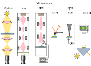 Types of microscopesMicroscopes can be separated into several different classes. One grouping is based on what interacts with the sample to generate the image, i.e., light (optical microscopes), electrons (electron microscopes) or a probe (scanning probe microscopes). Alternatively microscopes can be classed on whether they analyse the sample via a scanning point (confocal optical microscopes, scanning electron microscopes and scanning probe microscopes) or analyze the sample all at once (wide field optical microscope and transmission electron microscopes).
The wide field optical microscope and transmission electron microscope use the theory of lenses (optics for light microscopes and electromagnet lenses for electron microscopes) in order to magnify the image generated by the passage of a wave through the sample, or reflected by the sample. The waves used are electromagnetic (in optical microscopes) or electron beams (in electron microscopes). Resolution in these microscopes is limited by the wavelength of the radiation used to image the sample, shorter wavelengths allow a higher resolution.
Scanning optical and electron microscopes, like the confocal microscope and scanning electron microscope, use lenses to focus a spot of light/electrons onto the sample then analyze the reflected and/or transmitted waves. The point is then scanned over the sample to analyze a rectangular region. Magnification of the image is achieved by displaying the data from scanning a small sample area on a large screen. These microscopes have the same resolution limit as wide field optical and electron microscopes.
Scanning probe microscopes also analyze a single point in the sample and then scan the probe over a rectangular sample region to build up an image. As these microscopes do not use electromagnetic or electron radiation for imaging they are not subject to the same resolution limit as the optical and electron microscopes described above.
Sarfus, a recent optical technique increases the sensitivity of standard optical microscope to a point it becomes possible to directly visualize nanometric films (down to 0.3 nanometre) and isolated nano-objects (down to 2 nm-diameter). The technique is based on the use of non-reflecting substrates for cross-polarized reflected light microscopy.
 CBP Office of Field Operations agent checking the authenticity of a travel document at an international airport using a stereo microscopeUltraviolet light enables the resolution of microscopic features, as well as to image samples that are transparent to the eye. Near infrared light images circuitry embedded in bonded silicon devices, as silicon is transparent in this region. Many wavelengths of light, ranging from the ultraviolet to the visible are used to excite fluorescence emission from objects for viewing by eye or with sensitive cameras.
Phase contrast microscopy is an optical microscopy illumination technique in which small phase shifts in the light passing through a transparent specimen are converted into amplitude or contrast changes in the image. A phase contrast microscope does not require staining to view the slide. This microscope made it possible to study the cell cycle.
The traditional optical microscope has recently been modified into a digital microscope, where, instead of directly viewing the object, a charge-coupled device (CCD) is used to record the image, which is then displayed on a computer monitor.
[edit] The rise of modern light microscopy
See also: optical microscope
The first detailed account of the interior construction of living tissue based on the use of a microscope did not appear until 1644, in Giambattista Odierna's L'occhio della mosca, or The Fly's Eye.[3]It was not until the 1660s and 1670s that the microscope was used extensively for research in Italy, Holland and England. Marcelo Malpighi in Italy began the analysis of biological structures beginning with the lungs. Robert Hooke's Micrographia had a huge impact, largely because of its impressive illustrations. The greatest contribution came from Antoni van Leeuwenhoek who discovered red blood cells and spermatozoa and helped popularise microscopy as a technique. On 9 October 1676, Leeuwenhoek reported the discovery of micro-organisms.[3]
In 1893 August Köhler developed a key technique for sample illumination, Köhler illumination, which is central to modern light microscopy. This method of sample illumination gives rise to extremely even lighting and overcomes many limitations of older techniques of sample illumination. Further developments in sample illumination came from Fritz Zernike in 1953 and George Nomarski 1955 for their development of phase contrast and differential interference contrast illumination which allow imaging of transparent samples.
[edit] Electron microscopy

See also: electron microscope
In the early 1900s a significant alternative to light microscopy was developed, using electrons rather than light to generate the image. Ernst Ruska started development of the first electron microscope in 1931 which was the transmission electron microscope (TEM). The transmission electron microscope works on the same principle as an optical microscope but uses electrons in the place of light and electromagnets in the place of glass lenses. Use of electrons instead of light allows a much higher resolution.Development of the transmission electron microscope was quickly followed in 1935 by the development of the scanning electron microscope by Max Knoll.[4]
Electron microscopes quickly became popular following the Second World War. Ernst Ruska, working at Siemens developed the first commercial transmission electron microscope and major scientific conferences on electron microscopy started being held in the 1950s. In 1965 the first commercial scanning electron microscope was developed by Professor Sir Charles Oatley and his postgraduate student Gary Stewart and marketed by the Cambridge Instrument Company as the "Stereoscan".
[edit] Scanning probe microscopy
See also: scanning probe microscope
The 1980s saw the development of the first scanning probe microscopes. The first was the scanning tunneling microscope in 1981, developed by Gerd Binnig and Heinrich Rohrer. This was closely followed in 1986 with Gerd Binnig, Quate, and Gerber's invention of the atomic force microscope.[edit] Fluorescence and light microscopy
The most recent developments in light microscope largely centre on the rise of fluorescence microscopy in biology. During the last decades of the 20th century, particularly in the post-genomic era, many techniques for fluorescent labeling of cellular structures were developed. The main groups of techniques are small chemical staining of cellular structures, for example DAPI to label DNA, use of antibodies conjugated to fluorescent reporters, see immunofluorescence, and fluorescent proteins, such as green fluorescent protein. These techniques use these different fluorophores for analysis of cell structure at a molecular level in both live and fixed samples.The rise of fluorescence microscopy drove the development of a major modern microscope design, the confocal microscope. The principle was patented in 1957 by Marvin Minsky, although laser technology limited practical application of the technique. It was not until 1978 when Thomas and Christoph Cremer developed the first practical confocal laser scanning microscope and the technique rapidly gained popularity through the 1980s.
Main article: Microscopy#Sub-diffraction techniques
Much current research (in the early 21st century) on optical microscope techniques is focused on development of superresolution analysis of fluorescently labeled samples. Structured illumination can improve resolution by around two to four times and techniques like stimulated Emission Depletion microscopy are approaching the resolution of electron microscopes.[edit] Types

The wide field optical microscope and transmission electron microscope use the theory of lenses (optics for light microscopes and electromagnet lenses for electron microscopes) in order to magnify the image generated by the passage of a wave through the sample, or reflected by the sample. The waves used are electromagnetic (in optical microscopes) or electron beams (in electron microscopes). Resolution in these microscopes is limited by the wavelength of the radiation used to image the sample, shorter wavelengths allow a higher resolution.
Scanning optical and electron microscopes, like the confocal microscope and scanning electron microscope, use lenses to focus a spot of light/electrons onto the sample then analyze the reflected and/or transmitted waves. The point is then scanned over the sample to analyze a rectangular region. Magnification of the image is achieved by displaying the data from scanning a small sample area on a large screen. These microscopes have the same resolution limit as wide field optical and electron microscopes.
Scanning probe microscopes also analyze a single point in the sample and then scan the probe over a rectangular sample region to build up an image. As these microscopes do not use electromagnetic or electron radiation for imaging they are not subject to the same resolution limit as the optical and electron microscopes described above.
[edit] Optical
Main article: Optical microscope
The most common type of microscope—and the first invented—is the optical microscope. This is an optical instrument containing one or more lenses producing an enlarged image of a sample placed in the focal plane. Optical microscopes have refractive glass and occasionally of plastic or quartz, to focus light into the eye or another light detector. Mirror-based optical microscopes operate in the same manner. Typical magnification of a light microscope, assuming visible range light, is up to 1500x with a theoretical resolution limit of around 0.2 micrometres or 200 nanometres. Specialized techniques (e.g., scanning confocal microscopy, Vertico SMI) may exceed this magnification but the resolution is diffraction limited. The use of shorter wavelengths of light, such as the ultraviolet, is one way to improve the spatial resolution of the optical microscope, as are devices such as the near-field scanning optical microscope.Sarfus, a recent optical technique increases the sensitivity of standard optical microscope to a point it becomes possible to directly visualize nanometric films (down to 0.3 nanometre) and isolated nano-objects (down to 2 nm-diameter). The technique is based on the use of non-reflecting substrates for cross-polarized reflected light microscopy.

Phase contrast microscopy is an optical microscopy illumination technique in which small phase shifts in the light passing through a transparent specimen are converted into amplitude or contrast changes in the image. A phase contrast microscope does not require staining to view the slide. This microscope made it possible to study the cell cycle.
The traditional optical microscope has recently been modified into a digital microscope, where, instead of directly viewing the object, a charge-coupled device (CCD) is used to record the image, which is then displayed on a computer monitor.
Monday, March 7, 2011


Subscribe to:
Comments (Atom)